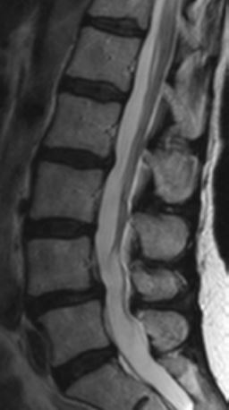
#HOW TO READ MRI IMAGES OF LUMBAR SPINE FREE#
If you require a scan with contrast feel free to get in touch and we’d be happy to recommend a trusted facility that can provide this service. Typically MRI of the lumbar spine is only performed with contrast if there is a suspected mass, infection, or recent lumbar surgery.Īt First Look MRI we only perform lumbar spine MRI scans without contrast. Adding contrast to an MRI scan significantly increases the cost so always check with your doctor to determine whether or not it is absolutely necessary. Though very rare, patients can be allergic to the contrast so it should only be administered if absolutely necessary. The contrast agent is injected into a vein in the arm and quickly circulates throughout the body, enhancing the areas of high blood flow. In a lumbar spine MRI with contrast, a contrast agent (a dye) is administered to highlight areas of increased blood flow or inflammation. Lumbar spine MRI with or without contrast?Ī lumbar spine MRI scans can be performed with or without contrast. A lumbar spine MRI helps your doctor determine the appropriate medical treatment for your pain which may be a rest, physical therapy, oral pain medicine, a pain management procedure such as an epidural steroid injection or nerve root block, or a surgical procedure such as a laminectomy and discectomy. Unexplained back pain with normal x-rays that does not get better with conservative treatmentĪ lumbar spine MRI scan can quickly and effectively identify the causes of low back pain and radiculopathy and rule out significant abnormalities resulting from work related or sports activities or accidents.Adenopathy (large lymph nodes) or masses around spine.Vertebral body infections (osteomyelitis).Congenital pars defects and pars fractures.Slippage or subluxation of lumbar spine segments (spondylolisthesis).Lumbar spine or sacral fractures which are not visible or hard to see on X-rays.Disc herniations (disc protrusions or extrusions).Degenerative disc disorders resulting in arthritis.MRI imaging of the lumbar spine is performed to diagnose: It allows a doctor to see if there are disc bulges or herniations, arthritis, injuries, tumors, or nerve abnormalities that may be causing the symptoms and require further attention. Unlike X-ray or CT scans which only visualizes the cortical bones and soft tissues, an MRI scan allows you to see marrow, spinal cord, nerve roots, discs, ligaments, muscles, and blood vessels.Ī lumbar spine MRI scan is a non-invasive medical exam that physicians use to assess low back pain, sciatica, lower extermity weakness, and shooting pain (radiculopathy) down one or both legs. No surgical incision is necessary and it can be performed on any part of the body. You'll be able to see this on a spinal MRI - there will be a long line of "normal" vertebrae and discs, with one noticeably bulging out.Lumbar spine MRI Scan What is a Lumbar Spine MRI Scan?Ī Magnetic Resonance Imaging (MRI) scan makes use of a combination of radio waves and magnets to capture images of the inside of the body. When you get a herniated disc, one of these discs breaks and the fluid leaks out, causing pain as it presses against the nerves in your spine. Between every two vertebrae is a fluid-filled disc. X Trustworthy Source Mayo Clinic Educational website from one of the world's leading hospitals Go to source The spine is made up of many different bone vertebrae stacked on top of each other. A good example of the second case is for spinal disc herniations.Similarly, for parts of the body that have many similar features repeated multiple times, a difference in one of the features can be a sign that something is amiss.
#HOW TO READ MRI IMAGES OF LUMBAR SPINE PATCH#
If, in your MRI, you notice a patch of lightness or darkness on one side of your body that does not match what's on the other side, this can be cause for concern. By and large, the body is very symmetrical.

Here, you're basically viewing thin slices of your body from the top down - as if you've been cut into many thin horizontal slices from your head to your toes like a salami.


When your MRI first loads up, if you're lucky, it will be immediately obvious what you're looking at. Familiarize yourself with the different MRI viewing schemes.


 0 kommentar(er)
